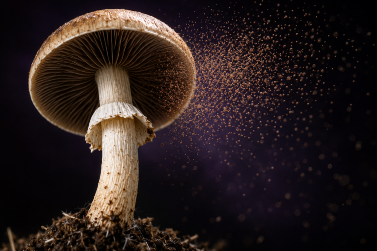
How to Identify Mushroom Spores Under a Microscope
Identify mushroom spores under microscope by examining their size, shape, colour and other microscopic characteristics. Learning how to identify mushroom spores under microscope is one of the most valuable skills in mycology because many species can only be distinguished through careful microscopic observation.
Learning to observe mushroom spores under a microscope allows beginners and experienced mycologists alike to better understand fungal biology and improve species identification.
Why Examine Mushroom Spores Under a Microscope?
Many mushroom species look remarkably similar in the field. Examining mushroom spores under a microscope provides additional information that cannot be seen with the naked eye.
Characteristics such as spore size, shape and surface texture are commonly used during fungal identification.
Equipment You’ll Need
To examine mushroom spores under a microscope, you’ll need:
- Compound microscope
- Clean microscope slides
- Cover slips
- Prepared spore print or spore sample
- Pipette or sterile water (if required)
A microscope capable of 400× magnification is generally suitable for observing many spore characteristics, while higher magnification provides additional detail.
Preparing a Microscope Slide
Begin by placing a very small amount of spores onto a clean microscope slide.
If using a dry spore print, a tiny sample is sufficient. Carefully position a cover slip over the specimen to create an even viewing surface.
Good slide preparation makes observing mushroom spores under a microscope much easier.
What Features Should You Look For?
Several important characteristics can be observed during microscopy.
Spore Size
Different mushroom species produce spores of different sizes. Measuring spores helps narrow identification.
Spore Shape
Spores may appear:
- Oval
- Elliptical
- Round
- Almond-shaped
- Elongated
Shape is one of the most useful identification features.
Spore Colour
Although individual spores are microscopic, their colour may become visible under appropriate lighting.
Combined with spore print colour, this information assists fungal identification.
Germ Pore
Some mushroom spores contain a small germ pore that can often be observed under higher magnification.
The presence or absence of a germ pore is another useful identifying characteristic.
Surface Texture
Some spores appear smooth while others display fine ornamentation.
These subtle differences are commonly used during advanced fungal identification.
Common Beginner Mistakes
When examining mushroom spores under a microscope, beginners often:
- Use too many spores on one slide
- Apply too much pressure to the cover slip
- Use poor lighting
- Attempt identification using only one characteristic
Observing several characteristics together usually produces the best results.
Frequently Asked Questions
What magnification is best?
Most observations begin around 400× magnification, although higher magnification may reveal additional detail.
Can all mushroom spores be identified under a microscope?
Microscopy provides valuable information, but accurate identification often requires examining several characteristics together.
Do all mushroom spores look the same?
No. Mushroom spores vary greatly in size, shape, colour and surface texture depending on the species.
Final Thoughts
Learning to examine mushroom spores under a microscope opens an entirely new perspective on fungal biology. By understanding spore size, shape, colour and microscopic features, mycology enthusiasts can gain a much deeper appreciation of mushroom diversity and identification.